

It's what's on the inside that matters
Dr. David Burk has very specific professional goals: make research easier and faster.
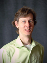
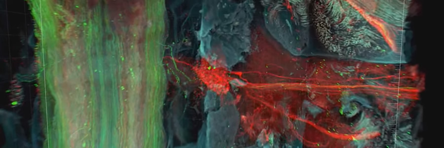
Burk runs the Imaging Core at Pennington Biomedical Research Center, where he helps scientists visualize and analyze information from their respective labs. Sometimes that work looks like science fiction as Burk renders a video that takes you inside the body to show how cells are functioning. This type of imaging is on the cutting edge of modern-day research.
These imaging techniques are allowing scientists to better envision the body's processes at the cellular level so they can lay the foundation for a better understanding of disease states. They then use this information to improve prevention and treatments for chronic diseases such as diabetes and obesity. These imaging methods are also faster than ever, allowing scientists more time in the lab and in the office for research.
"We are on par with some of the top research institutions in the nation," said Burk. "In the Imaging Core, our goal is to reveal things that we weren't able to see previously, all in an effort to ultimately help people achieve and maintain better health."
For more information on how you can support this and other projects at LSU’s Pennington Biomedical Research Center, visit www.pbrf.org.
